
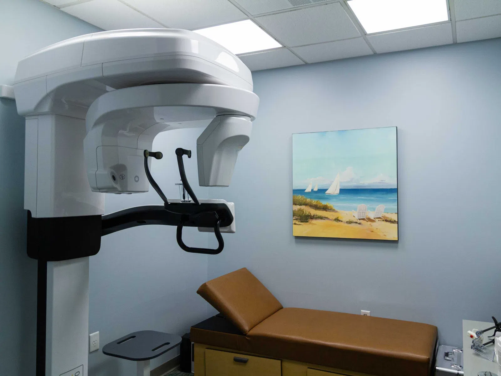
Cone Beam CT
Benefit From Modern Surgical Care
Advanced Images From Our Cone Beam CT in Virginia Beach Help Plan Your Surgery
A 3-D view of your jaw helps your surgery go smoothly and lowers the chance of surprises. Our team uses a cone beam CT in Virginia Beach to see your jaw structure in far greater detail than standard X-rays can show. The scan takes less than a minute while you sit or stand comfortably. It reveals bone height and width along with the location of nerves and sinuses. That information helps us plan your dental implant placement, including guided procedures. It also helps our surgeons map impacted wisdom teeth and canines before treatment.
Cone beam imaging uses less radiation than a medical CT scan, giving you more peace of mind. Schedule a consultation to learn how we use 3-D imaging to shape your treatment plan.
Discover Why Cone Beam 3-D Imaging Matters
Standard dental X-rays show teeth and bone in flat, two-dimensional images. Three-dimensional CT imaging goes further. A cone-shaped X-ray beam rotates around your head and captures hundreds of images in a single pass. Software then combines them into a 3-D model that your surgeon can view from any angle.
This model shows details that flat images can miss, such as bone thickness and the exact path of the jaw nerve. Our surgeons use these scans when planning implants and bone grafts. They also rely on this detailed imaging to evaluate jaw cysts and impacted teeth before surgery.
Better information leads to better planning. With a clear picture, we can choose the safest path and explain your options with confidence. Your surgeon can also walk you through the images to make your treatment easier to understand.
See how our cone beam CT in Virginia Beach can improve your treatment. Contact us today for a consultation.
Complete 3-D Detail
A cone beam scan shows your jaw from every angle, revealing details that flat X-rays often miss.
Safer Implant Planning
Precise bone measurements help your surgeon choose the right implant size and keep clear of nearby nerves.
Quick, Comfortable Scan
The scan takes less than a minute, and you simply sit or stand still while the machine rotates.
Lower Radiation Dose
Cone beam CT scanning uses far less radiation than a hospital CT scan while still giving detailed images.
Guided Surgery Ready
Your scan becomes the foundation for a guided surgery procedure, linking careful planning to precise placement.
Frequently Asked Questions About Cone Beam CT
Q What is cone beam CT?
Cone beam CT is a type of 3-D dental imaging that captures detailed views of the jaws and surrounding structures. A cone-shaped X-ray beam rotates around the head in a single sweep. Software turns the images into a 3-D model that the surgeon can study from any angle.
Q How is cone beam CT imaging different from a regular dental X-ray?
Q How much radiation does a cone beam CT scan use?
A cone beam CT scan uses a low dose of radiation, typically much lower than a medical CT scan. The dose varies based on the size of the area scanned. Smaller scans focused on one section of the jaw use even less. Providers take scans only when they are clinically useful.
Q How long does a cone beam CT scan take?
A cone beam CT scan usually takes less than a minute. The person sits or stands while the machine rotates around the head. Staying still during the scan helps produce clear images. The whole appointment, including positioning, often takes only a few minutes. Results are ready for review almost immediately.
Q Does a cone beam CT scan hurt?
A cone beam CT scan does not hurt. The machine moves around the head without causing discomfort, and there are no needles. Most people simply bite gently on a small positioner and hold still for a few seconds. People who feel anxious in tight spaces usually find the open design comfortable.
Q Why do oral surgeons use a cone beam CT before dental implants?
Oral surgeons use a cone beam CT before dental implants to measure bone height and width precisely. The scan shows nerves and sinuses that implants must avoid. This information helps the surgeon choose the right implant size and position. It also provides the data that guides robotic and navigation systems during placement.
Q Can cone beam CT scanning help with wisdom teeth removal?
Cone beam CT scanning can help with wisdom teeth removal by showing the exact position of each tooth and its roots. It reveals how close the roots sit to the nerve in the lower jaw. This helps the surgeon plan a safer extraction and lower the risk of nerve injury.
Q Is cone beam CT imaging safe for children and teens?
Cone beam CT imaging is considered safe for children and teens. Providers use the smallest scan area and lowest dose possible for young people. It is often used to locate impacted canines or extra teeth.

